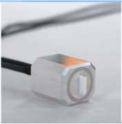
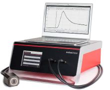
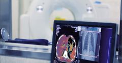
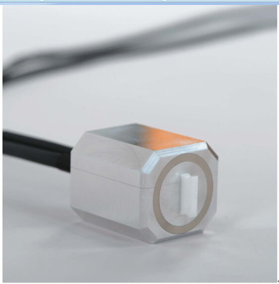
 产品介绍 一、产品介绍: 该系统适用于药物动力学血液放射活度实时测量研究(可配合于PET、SPECT、PET/MRI系统) Twilite 是由 Swisstrace 公司所研发设计的高灵敏度自动血液取样系统。此系统可与 PET 、SPECT、或 PET/MR 影像系统结合使用,无论是小至实验动物、大至其他更大的个体,均能够在线高分辨率采集血液活度实时变化数据。 Twilite 系统的核心是一个设计精巧的侦测头(探测器),由 LYSO 晶体与屏蔽外来辐射用的医疗级钨加工製成,因此完全与 MR 影像系统相容。闪烁信号透过两条可自订长度的高效率光导管传输至光子侦测单元。此设计的侦测头端完全没有任何电子零件,所以能够避免来自其他设备所造成的电磁干扰问题。此外,这样的设计也能够将人体研究实验的潜在风险最小化。 数据采集是使用 PMOD 公司所开发的 PSAMPLE 软件,藉由 TCP/IP 介面传输,允许同时记录多套 Swisstrace 系统的讯号,例如可同时使用 Twilite 系统与 Twin beta probe 系统。此外,尚有两个类比讯号输入孔可同时记录来自其他仪器的讯号,例如Laser Doppler Flowprobes、ECG 或来自辅助设备的触发讯号。 PMOD 软件的功能模块可对取得的放射活度信号进行离线处理分析。 此系统也脱离计算机独立工作。仪器前方的触摸式面板可直接进行操作,并即时显示测量数据。 Twilite 系统性能优越。除了拥有**的灵敏度外,即使在高辐射值的环境下,仍然呈现出稳定的线性度与信噪比。 Swisstrace 公司的开发人员在放射定量实验方面具有相当深厚的经验。系统设计乃针对 PET 系统(包含小动物与人)**化。侦测头精巧的尺寸尤其适合使用于小动物正子造影系统中,搭配动、静脉分流管(arterio-venous shunt), Twilite 系统可测得全血的动脉输入函数(arterial input function, AIF)而不必将血液抽离体外。 二、仪器结构组成(1-9项为产品标配): 图1 图2 图3 1、连接股动脉与股静脉的分流管 (自购) 2、蠕动帮浦(Peristaltic Pump)(自购) 3、Twilite 钨制探测器 4、LYSO 晶体1 5、LYSO 晶体2 6、光导管:传输光子讯号至PMT。标准长度2 m,若需使用于MR 系统可延长至10 m 7、光子侦测单元 8、两个模拟讯号输入孔(可与其他品牌仪器配合使用,监控呼吸、ECG 或血压等) 9、TCP/IP 传输接口:可透过因特网传输或直接与计算机连接,使用PMOD 软件PSAMPLE 模块进行数据采集 10、安装PMOD 软件的计算机,进行数据采集与分析(自购) 结构说明:动静脉分流管(小鼠用PE10,大鼠用PE50)可同时用于血压量测、药物注射及血液样本采集等其他功能,如图3所示。血液样本采集可用解剖刀在导管上划一个小口,在采集时间点将导管往缺口方向推,即可取得血液样本。 ●结构与曲线函数(如下图) 左图为实验架构。血流以蠕动泵驱动,从股动脉流出体外,经过耦合讯号侦测头后,再由股静脉回到体内。t1与t2两个三向阀分别用来进行血液取样与药物注射。右图为Twilite 系统所测得的小鼠动脉输入曲线。 三、系统规格: 四、用户名单: 五、合作伙伴: PMOD Technologies Ltd. Unitectra Zurich, Switzerland Zurich, Switzerland University of Zurich CSEM Zurich, Switzerland Neuch?tel, Switzerland 六、药物动力学实验论文(部分摘要): Quantification of Brain Glucose Metabolism by 18F-FDG PET with Real-Time Arterial and Image-Derived Input Function in Mice Malte F. Alf1,2, Matthias T. Wyss3,4, Alfred Buck3, Bruno Weber4, Roger Schibli1,5, and Stefanie D. Kr?mer11Center for Radiopharmaceutical Sciences of ETH, PSI, and USZ, Institute of Pharmaceutical Sciences, Department of Chemistry and Applied Biosciences, ETH Zurich, Zurich, Switzerland; 2Laboratory of Functional and Metabolic Imaging, Institute of Physics of Biological Systems, Ecole Polytechnique Fédérale de Lausanne, Lausanne, Switzerland; 3Department of Nuclear Medicine, University Hospital Zurich, Zurich, Switzerland; 4Institute of Pharmacology and Toxicology, University of Zurich, Zurich,Switzerland; and 5Center for Radiopharmaceutical Sciences of ETH, PSI, and USZ, Paul Scherrer Institute PSI, Villigen, Switzerla Kinetic modeling of PET data derived from mouse modelsremains hampered by thetechnical inaccessibility of an accurateinput function (IF). In this work, we tested the feasibility of IF measurement with an arteriovenous shunt and a coincidencecounter in mice and compared the method with an imagederived IF (IDIF) obtained by ensemble-learning independent component analysis of the heart region. Methods: 18F-FDG brain kinetics were quantified in 2 mouse strains, CD1 and C57BL/6, using the standard 2-tissue-compartment model. Fits obtained with the 2 IFs were compared regarding their goodness of fit as assessed by the residuals, fit parameter SD, and Bland–Altman analysis. Results: On average, cerebral glucose metabolic rate was 10% higher for IDIF-based quantification.The precision of model parameter fitting was significantly higher using the shunt-based IF, rendering the quantification of single process rate constants feasible. Conclusion: We demonstrated that the arterial IF can be measured in mice with a femoral arteriovenous shunt. This technique resulted in higher precision for kinetic modeling parameters than did use of the IDIF. However,for longitudinal or high-throughput studies, the use of a minimally invasive IDIF based on ensemble-learning independent component analysis represents a suitable alternative. Key Words: energy metabolism; PET; molecular imaging; glucose; kinetic modeling J Nucl Med 2013; 54:1–7 DOI: 10.2967/jnumed.112.107474 PET with 18F-FDG is a commonly used method to determine glucose metabolism in animal and human tissues (1). Full quantification of 18F-FDG kinetics can be achieved by applying a 2-tissue-compartment model (2). The model requires the time course of the 18F-FDG concentration in the target organ(tissue time–activity curve) and in arterial plasma (input function, IF). In human brain PET, the IF is commonly measured from a catheter placed in the radial artery. An alternative is derivation of the IF from PET images via values measured in a volume of interest placed over the cardiac ventricles or a large vessel. A prerequisite of image-derived IFs (IDIFs) is the location of the blood pool and the organ of interest in the same field of view. In general, one or more arterial blood samples are measured to calibrate the IDIF. In a recent review article for human PET(3), the authors concluded that arterial blood sampling remains the preferred method to define the IF, because invasiveness is hardly reduced by the use of an IDIF. In small animals, the small blood volume is the major constraint for manual blood sampling. This constraint prompted the development of several alternative methods, such as the sampling of very small volumes via a microfluidic chip (4) or the use of b-probes for measuring the blood radioactivity (5,6). Despite these new physical methods, the main research focus has been on developing the use of IDIFs, where blood radioactivity is estimated directly from the dynamic PET images. IDIF generation from simple analysis of blood pool volumes such as the liver or the heart ventricles is flawed by 18F-FDG uptake by the liver or spillover from surrounding myocardium, cardiac motion, and partial-volume effects. Compensation can be achieved to varying degrees by image processing methods such as factor analysis (7), modelbased techniques (8), independent component analysis (9), so-called hybrid IDIFs (e.g., 10,11), and cardiac gating combined with improved image reconstruction algorithms (12). Most of these methods rely on at least 1 measure from a blood sample for scaling of the IDIF.Hence, blood sampling is not entirely obviated. To our knowledge, there is currently no gold standard to define the real-time 18F-FDG arterial IF in mice in a reliable and easily accessible manner. In this study, we adapted a method for direct blood radioactivity measurements and an approach for the generation of IDIFs for use in mice. We acquired real-time blood radioactivity curves by means of a new coincidence counter in combination with an arteriovenous shunt and compared the findings to IDIFs generated from PET data of the cardiac region with an ensemblelearning independent component analysis (EL-ICA) algorithm (13).We used 2 different mouse strains to explore the possible strain dependency of our methods: C57BL/6 mice, because they are relevant for studies of genetically modified animals, and CD1 mice, because they are valuable as disease models,such as cerebral ischemia (14). The purpose of this work was 2-fold. First, we evaluated whether the arteriovenous-shunt/ counter technique, which was previously demonstrated in rats (15), is also feasible in mice. Second, we compared 18F-FDG kinetic parameters and fit precisions obtained with the experimental shunt IF and the IDIF. |
相关产品
|